
A yellow plaque sits at the inner corner of the upper eyelid, and two clinics give two different answers. One offers xanthelasma laser treatment with no stitches; the other recommends cutting the plaque out. Both can be right, for different plaques. Laser and surgical excision suit different lesions, and the choice turns on how thick the plaque is, where it sits on the lid, how many there are and how much spare eyelid skin is available.
This guide sets out that decision the way it is made at a consultation: a plaque-by-plaque decision matrix, then the practical differences in sessions, stitches, downtime and the mark each method tends to leave, including pigment change on Indian skin. How the CO2 laser works and the week-by-week recovery are covered on the xanthelasma treatment page.
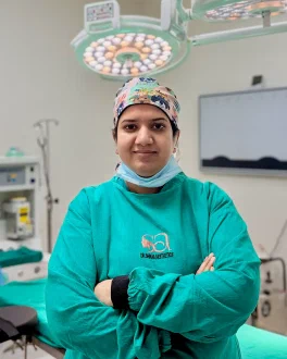
In her practice in Gurgaon, Dr. Shikha finds the method is often clear once the lid is examined in good light. The longer conversation is usually about which trade-off sits better with the patient: a fine line, or a patch that may change colour.
Who this article is for
- Anyone offered laser at one clinic and excision at another, wanting to understand why
- Anyone not yet sure the bumps are xanthelasma: milia and syringoma look similar and are treated differently
- Anyone with hooded upper lids wondering whether the plaque and the loose skin can be dealt with together
- Anyone with medium-to-dark skin worried about a treated patch healing darker or lighter
- Anyone whose xanthelasma returned after an earlier laser or chemical treatment
Laser or surgery for xanthelasma: which suits your plaque?
For flat, thin plaques in the upper layers of the skin, laser treatment for xanthelasma is usually the first choice: no stitches and, in most cases, a single session. For thick or large plaques (roughly over 10 mm), surgical excision tends to clear the deposit more reliably in one sitting, leaving a fine scar in the eyelid crease. When the upper lid also has excess skin, excision can often be combined with a blepharoplasty-style skin removal. The plaque decides the method, not a clinic’s preference for one machine.
The matrix below shows how each feature tends to push the decision. Most plaques have features pointing both ways, which is why the final call is made in person.
| Plaque feature | Tends to favour laser | Tends to favour excision |
|---|---|---|
| Thickness | Flat, thin, superficial | Raised, thick, deeper in the skin |
| Size | Small, under about 10 mm | Over about 10 mm, or merging plaques |
| Number | Several small, scattered plaques | One or two large, defined plaques |
| Eyelid skin | Tight lid skin with little to spare | Loose or hooded upper lid skin |
| Pigment risk | Small treatment area | Large area on darker skin |
| History | First treatment of a thin plaque | Recurrence after laser or chemical treatment |
Radiofrequency and TCA chemical cauterisation are also used for some thin plaques; both are described on the procedure page.
How each plaque feature shifts the decision
Thickness matters most, then location on the lid, then how much skin can safely be removed. Laser removes tissue layer by layer from the surface, so the deeper the plaque, the deeper the treatment goes and the higher the chance of a dent or colour change. Excision removes the full depth at once but uses up eyelid skin, and the eyelid has only so much to spare before it starts to pull.
Flat versus raised or thick plaques
A flat plaque that barely lifts the skin usually suits laser. Clearing a raised or thick plaque fully with laser risks a depressed or lighter patch, so it is either staged across sessions or excised. Leaving a thin layer behind to avoid a mark is a common reason plaques return early.
Upper lid, lower lid and the inner corner
Most xanthelasma begins at the inner corner of the upper lid, where the crease offers a natural place to hide an excision line. The lower lid has less spare skin, and removing too much can pull the lid margin away from the eye (ectropion), so lower-lid excision is planned conservatively. At the inner corner, the tear drainage channel runs close to the lid margin, which limits how wide an excision can go; large plaques here occasionally need a small local flap to close.
One plaque or many
Several small, scattered plaques can often be lasered in one sitting. Plaques on all four lids, or plaques merged into a band, are usually staged: upper lids first, lower lids once those have healed. Excising widespread plaques all at once risks over-tightening the lids so the eyes do not close fully (lagophthalmos).
Loose upper lid skin
When the upper lid is already hooded, a plaque within the fold can sometimes be removed as part of the skin taken in a blepharoplasty, so one crease scar deals with both concerns. On tight skin, the same width of excision carries more risk of the lid pulling.
Pigment risk on Indian skin
On medium-to-dark Indian skin (Fitzpatrick IV to V), a lasered patch commonly heals darker than the surrounding lid skin and usually fades over months, occasionally a year or more. A lighter patch is less common but can be permanent. The larger the area lasered, the larger the area exposed to that risk, so for a large plaque on darker skin, excision trades a patch for a fine line.
Does laser treatment for xanthelasma need more than one session?
A flat, thin plaque is usually cleared in one laser session. A thicker plaque may need two sessions spaced several weeks apart, to avoid going too deep at once. Surgical excision is typically a single sitting per area, though plaques on both upper and lower lids are often staged. Both are day procedures under local anaesthesia.
Stitches and downtime
Laser leaves no stitches; a small scab forms and separates on its own over about a week to ten days. Excision is closed with fine sutures, usually removed around day five to seven. Swelling and bruising tend to be more noticeable after excision, while the laser site stays pink for longer. Desk work is commonly comfortable within a few days of either.
What laser surgery for xanthelasma cannot do
Laser cannot remove excess skin. Neither method stops new plaques forming. Xanthelasma can be the first visible sign of raised cholesterol, although around half of people with it have normal lipid levels, so a fasting lipid profile and a check with your physician are usually advised alongside removal. Managing any lipid problem is good for your overall health, though plaques can still return even when cholesterol is normal.
Scar or colour change: what each method leaves behind
Excision tends to leave a fine line in the eyelid crease that is pink and slightly firm for the first weeks and usually settles over three to six months. Laser tends to leave no line but can leave a slight texture change or a patch of altered colour, particularly on darker skin. Neither method leaves an invisible result; the better method is the one whose likely mark is easier to live with.
A small depression at the excision site occurs in a minority of cases and often improves as the scar matures. After laser, a shallow dent can follow deep treatment. The same line-versus-patch trade-off applies to mole removal by surgery or laser.
Xanthelasma can come back after either method; the comparison is in why xanthelasma comes back after removal.
Planning xanthelasma removal in Gurgaon
The fee depends mainly on the method and how many plaques are treated, and rises when treatment is staged. A single small plaque treated with laser is a smaller procedure than a staged excision across four lids or an excision combined with a blepharoplasty, so a figure is given after examination rather than over the phone.
Laser for a small plaque can sometimes follow the consultation on the same day. Excisions are booked separately, with a follow-up around one week for suture removal or scab review.
Frequently asked questions
Is laser better than surgery for xanthelasma removal? Neither is better across the board. Laser tends to suit flat, thin, small plaques; excision tends to suit thick or large ones and plaques that have come back.
Can CO2 laser treatment for xanthelasma remove thick plaques? It can, but usually over more than one session, and deeper treatment raises the chance of a dent or colour change. For a thick or large plaque, excision is often more predictable.
Will xanthelasma laser removal leave a dark mark on Indian skin? A darker patch is common after laser on medium-to-dark skin and usually fades over several months. A lighter patch is less common but can be permanent, so this is discussed before a laser session is booked.
Is laser treatment for xanthelasma cheaper than excision? For a single small plaque, laser is usually the smaller procedure. Fees rise with the number of plaques, staging, and whether excision is combined with eyelid surgery, so a reliable quote follows examination.
Can xanthelasma be removed during eyelid surgery? Yes, when the plaque sits within the upper-lid skin a blepharoplasty would remove anyway. Both concerns are then handled with one crease scar.
Choosing between laser and excision is less about which technology sounds newer and more about which likely mark and recovery fit a particular plaque. Dr. Shikha Bansal (MBBS, MS General Surgery, MCh Plastic & Reconstructive Surgery; Haryana Medical Council Reg No. 24859) assesses plaque depth, lid position, skin laxity and skin tone together before recommending a method. This article is general information only and is not a substitute for medical advice; consult a qualified plastic surgeon about your own eyelids. Book a consultation