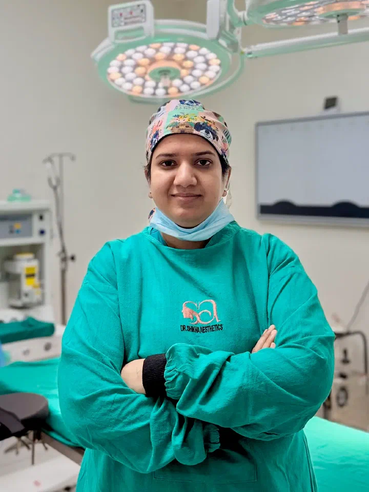

A lipoma is a soft, slow-growing lump of mature fat that sits just under the skin - usually painless, movable under the fingers, and benign. Lipoma removal at Dr. Shikha Bansal’s clinic in Gurgaon is done by surgical excision, or by liposuction-assisted removal where the lump is large and scar length matters more than the specimen. Most lipomas come out under local anaesthesia as a day-care procedure with no overnight stay. Every specimen removed is sent for histopathology, so the diagnosis is confirmed on a report rather than assumed from the examination.
Key points
- Every specimen removed is sent for histopathology, with the report explained and handed over
- Local anaesthesia and same-day discharge for most subcutaneous lipomas, with no overnight stay
- Incision planned for the body site - forehead crease, hairline, neck crease, or along skin tension lines
- Clinical examination first, with ultrasound or MRI arranged when the lump is large, deep or atypical
- Forehead, scalp and facial lipomas managed by a plastic surgeon where the lump is a straightforward lipoma
- Multiple lipomas planned in staged sittings, with the number per sitting agreed in advance
What a lipoma is, and how it differs from a sebaceous cyst or other lump
A lipoma is a benign tumour of mature fat cells in a thin capsule, sitting between the skin and the muscle. It is one of the commonest soft-tissue lumps in adults, and it is not cancer. Lipomas appear most often on the back, shoulders, neck, upper arms, chest wall and thighs, and - less often but far more visibly - on the forehead and scalp.
Lipoma or sebaceous cyst? A sebaceous (epidermoid) cyst sits within the skin rather than under it, so the skin moves with the lump instead of over it. It feels firmer, often has a small central punctum, and can turn red, tender and swollen if it gets infected. Lipomas do none of that. Other lumps mistaken for one include an enlarged lymph node, a ganglion, a dermatofibroma and an abscess, and a lump in the armpit has its own differential - axillary breast tissue is often mistaken for an armpit lipoma.
Examination settles the question in most cases. These features place a lump in the straightforward-lipoma category:
- Soft or doughy rather than firm or hard.
- Mobile - it slips sideways under the fingertip instead of staying fixed.
- Painless in most cases, though some ache when they sit under a bra strap, a waistband, or on a nerve.
- Slow-growing over months to years, not over weeks.
- Normal skin over it: no punctum, no redness, no discharge, no ulceration.
- Usually 1 cm to 5 cm across, though larger ones do occur.
When a lump is not a simple lipoma - red flags, and how one is diagnosed
Almost all fatty lumps are exactly what they look like. A small number are not, and those tend to announce themselves.
Clinical examination first. The lump is measured, its consistency and mobility assessed, the overlying skin checked, and the area examined for other lumps. For a small, soft, mobile, painless swelling in the subcutaneous layer with none of the red flags below, that is usually enough to plan an excision.
Ultrasound when the picture is not clean. Quick, no radiation, and it answers the useful questions - is the lump fatty, how deep does it go, one lesion or several, above or below the muscle fascia.
MRI when the lump is large, deep or atypical. Reserved for lumps over roughly 5 cm, lumps deep to the fascia or between muscles, awkward anatomical sites, and equivocal ultrasounds, because it shows the relationship to nerves and vessels.
Where imaging suggests the lump is not a simple lipoma, the honest step is a referral for a full soft-tissue work-up rather than a day-care excision - a call made before the patient is booked, not discovered on the table. The checklist below is what moves a lump into imaging first:
- Rapid growth - visible change over weeks rather than years.
- Larger than about 5 cm, particularly on the thigh, buttock or shoulder girdle.
- Firm, hard or rubbery rather than soft and doughy.
- Fixed - it does not glide under the skin, or the skin cannot be lifted off it.
- Deep to the muscle fascia rather than in the fat layer just under the skin.
- Painful at rest, or with numbness, tingling or weakness beyond it.
- A lump that has come back at the site of a previous removal.
- Any change in the overlying skin, or a new soft-tissue lump in someone with a past history of cancer.
Three routes for a lipoma: watchful waiting, surgical excision, liposuction-assisted removal
Not every lipoma needs an operation. The three routes are laid out side by side at consultation.
Watchful waiting suits a small, soft, mobile, painless lipoma with a typical examination and no red flags. The lump is measured and photographed for a baseline, and the patient returns if it grows or changes. A lipoma that is not bothering anyone does not have to be removed.
Surgical excision is the definitive route, and the one most patients choose once the lump is visible, catching on clothing, or simply unwanted. An incision is made over the lump, the capsule is dissected free, and the lipoma is delivered whole. Because the capsule comes out with it, recurrence at that site is uncommon, and the intact specimen is what a pathologist needs for a clean report.
Liposuction-assisted removal uses a small incision, often sited away from the lump, and a cannula to break up and aspirate the fat. It is worth considering for large, soft lipomas on the trunk or back where a conventional excision would leave a long scar. The trade-off is stated plainly: part of the capsule tends to remain, making recurrence more likely, and the aspirate is fragmented, so it is less useful for histopathology. It is offered only where examination and imaging have confirmed a simple benign lipoma - any lump carrying a red flag is excised whole so the pathologist receives an intact specimen. The liposuction page covers the cannula work in more detail.
Talk it through with Dr. Shikha
Book a consultation at Dr. Shikha Aesthetics in Sushant Lok 1. Bring your questions about lipoma removal, and any previous reports.
Lipoma treatment without surgery - what works, and what does not
Searches for lipoma treatment without surgery, injection for lipoma removal, and creams that dissolve a lipoma return a great deal of confident marketing. The honest position is narrower.
No cream, oil, spray, tablet or home remedy has been shown to dissolve a lipoma. A lipoma is encapsulated fat under the skin; a topical product does not reach it, and nothing applied to the skin surface removes a capsule. Ayurvedic and homeopathic preparations marketed for lipoma have not been shown in controlled studies to remove one, and none of these are offered at the clinic.
Steroid injection can shrink some small, superficial lesions to a degree, but this is an off-label use supported only by small series, the effect is inconsistent, and it leaves the capsule behind. Injectable deoxycholate has been reported in small studies and remains investigational for this indication. Neither produces a specimen for histopathology, and neither is offered as a routine alternative to excision.
What matters clinically is that trying a remedy does not delay assessment of a lump carrying any red flag.
Why every lipoma removed at the clinic is sent for histopathology
“Is this cancer?” is the question most patients arrive with, and it deserves an answer on paper rather than reassurance across a desk.
Every lump excised at the clinic goes for histopathological examination as a routine, not as an optional extra the patient has to ask for. The pathologist confirms the tissue is a lipoma, identifies the variant where relevant - angiolipoma, fibrolipoma and spindle cell lipoma behave slightly differently - and, most importantly, excludes a liposarcoma. A liposarcoma is an uncommon malignant fatty tumour, but it can look and feel like a large lipoma, particularly when the lump is deep, over 5 cm, or on the thigh. Examination alone cannot separate the two with certainty in every case. Microscopy - with molecular testing where the picture is borderline - is what separates them.
The report typically comes back in about a week, is explained to the patient, and a copy is handed over. Where a report is atypical, the referral to a surgical oncology service is made from the clinic rather than left to the patient to chase.
Scar planning by body site - and why forehead and scalp lipomas are a separate case
The scar is the part of a lipoma removal the patient lives with, and the part most often left unplanned. A lipoma can usually be taken out through an incision shorter than the diameter of the lump, because fat delivers through a small opening once the capsule is freed.
Back, shoulder and chest wall carry among the highest skin tension on the body, and scars there tend to widen or thicken more than elsewhere. The incision is aligned with the relaxed skin tension lines and closed in layers so the deep layer takes tension off the skin. A personal or family history of keloid scarring is flagged at consultation and managed on the protocol set out on the keloid treatment page. Arm, forearm and thigh are more forgiving - the incision follows the long axis of the limb or a skin crease. Neck incisions go in a horizontal crease so the scar sits in a shadow line.
Forehead and scalp lipomas are the reason a plastic surgeon is often the right operator for what sounds like a minor lump. Forehead lipomas frequently sit deep to the frontalis muscle, close to the supraorbital nerve, so the dissection plane matters and the approach is planned to protect sensation. The incision goes in a forehead crease, at the hairline, or within hair-bearing scalp, angled parallel to the hair follicles so hair grows back through the line. Where an earlier removal elsewhere left a wide or thickened scar, the options are on the scar revision page.
The procedure, from consultation to same-day discharge
The first consultation runs about 20 to 30 minutes: the lump is examined and measured, other lumps are looked for, photographs are taken, and the incision is discussed against the specific site in the mirror. Blood thinners, diabetes, previous excisions and any keloid history are noted, and imaging is arranged at this visit if the checklist calls for it.
Most excisions are day-care procedures under local anaesthesia. Lignocaine with adrenaline is infiltrated around the lump; once the area is numb the incision is made, the capsule is separated from surrounding tissue, and the lipoma is lifted out whole. The cavity is checked for bleeding, closed in layers and dressed. A single subcutaneous lipoma usually takes 20 to 45 minutes. Large lipomas, lumps deep to the muscle fascia, and cases where several are removed together may need sedation or general anaesthesia in a day-care operating room - decided before the day rather than on it. In almost every case the patient goes home the same day; an overnight stay is occasionally advised, for example after a large or deep excision under general anaesthesia, or where the patient’s health calls for it.
It is a small operation, and it is still an operation. The risks are stated before consent: bleeding or a collection of blood under the wound, a seroma where a large lipoma leaves a cavity, infection, a scar that widens or thickens, numbness around the scar that usually settles over months, a contour dip where a bulky lipoma was removed, injury to a nerve or vessel running close to the lump, a reaction to the local anaesthetic (or the general risks of sedation or general anaesthesia where these are used), recurrence, and the possibility of further surgery if the histopathology report comes back atypical.
Patients come from across Delhi NCR - Gurgaon, Delhi, Noida, Faridabad and Ghaziabad - and consultations are by appointment. For a small, typical subcutaneous lipoma with no red flags, the consultation and the excision can sometimes happen on the same day when the schedule allows; lumps needing imaging and multiple-lipoma cases are booked separately so the scan is reviewed before anything is planned.
Recovery after lipoma removal, week by week
Recovery is short and predictable for a subcutaneous lipoma. Deep or very large lesions run roughly a week longer at each stage.
Day 0 to Day 2: mild soreness, usually managed with paracetamol, and the dressing kept dry. Where a large cavity was left, a compression dressing reduces the chance of a seroma.
Day 3 to Day 7: soreness settles and most patients are back at desk work within one to three days. The first dressing change and wound check is at day five to seven.
Suture removal, day 5 to day 14: sutures come out around day five to seven on the face and neck, day ten to fourteen on the trunk and limbs. The histopathology report is usually ready in this window and is discussed at the same visit.
Week 2 to Week 4: light activity resumes from week two, while gym work, lifting and swimming are held back until about week three to four for trunk, back and shoulder sites, since early tension there widens a scar. Silicone gel or paper tape starts once the wound is fully closed.
Month 2 to Month 6: the scar is pink and firm at first, then softens and fades toward the surrounding skin tone, with most of the fading happening between month three and month six, though a scar continues to mature and soften for 12 to 18 months.
Multiple lipomas, lipomatosis, and whether a lipoma can come back
Some patients have one lipoma. Others have ten or thirty, often with a parent or sibling showing the same pattern - familial multiple lipomatosis. Dercum’s disease and Madelung’s disease also present with multiple fatty swellings, and where the pattern suggests one of them a physician work-up is arranged alongside the surgical plan rather than after it.
How many can be removed in one sitting is limited by the total dose of local anaesthetic that can safely be given, how far apart the lumps sit, and how long the patient is comfortable in one position. A handful of small subcutaneous lipomas in the same region is a realistic single sitting under local anaesthesia; larger numbers, or lumps spread across the back, arms and thighs, are usually staged across visits or done in fewer sittings under sedation.
Recurrence at the same site is uncommon after a complete excision that removes the capsule intact, and is reported more often where a lipoma was removed piecemeal or by liposuction and part of the capsule was left behind. A new lipoma elsewhere is not a recurrence - in multiple lipomatosis, new lumps over the years are expected, and each is assessed on its own merits.