

Breast reduction surgery, or reduction mammoplasty, reduces heavy breasts while reshaping and lifting them into a lighter, more proportionate position. Breast reduction at Dr. Shikha Bansal’s clinic in Gurgaon is planned for back, neck, shoulder, and posture strain, bra-strap grooves, under-breast rashes, activity restriction, and aesthetic imbalance. Two separate choices are made rather than one: the pedicle that keeps the nipple-areola complex alive - inferior, superomedial, or superior - and the skin pattern that sets the scars, either a vertical lollipop or a Wise-pattern anchor. Most anchor-pattern reductions use an inferior pedicle as well, so those are not alternatives to each other; liposuction-only reduction is the separate non-excisional option.
Key points
- Symptom-led assessment - back, shoulder, and posture strain documented, not cup size alone
- Pedicle and skin pattern decided as two separate choices, with the anchor-pattern in-fold scar named upfront
- Scar, nipple-sensation, and breastfeeding trade-offs stated before the technique is chosen
- Pedicle and scar pattern chosen by breast size, ptosis, skin quality, tissue type, and goal
- Reduction planned with the lift component included, because the nipple is repositioned and the mound reshaped
- Recovery mapped from Week 0 to Month 3, with support-bra, work, and exercise milestones
- Durability framed around weight stability, pregnancy, scars, and revision triggers
When breast reduction is medical relief, not only a size change
Breast reduction is chosen when breast weight has become a physical problem, not just when the patient wants a smaller cup size. Heavy breasts can pull the shoulders forward, deepen bra-strap grooves, worsen upper-back and neck pain, cause sweating or intertrigo under the breast fold, and make exercise or fitted clothing difficult.
The same operation removes excess gland, fat, and skin, lifts the nipple-areola complex, narrows the lower pole, and reshapes the mound. The aim is a lighter breast that still looks natural for the patient’s frame.
A reduction may be symptom-led, aesthetic-led, or both. At consultation, Dr. Shikha Bansal documents symptoms, measurements, ptosis, skin quality, areola size, asymmetry, weight pattern, and future pregnancy or breastfeeding plans. A patient seeking relief from pain may accept a larger scar for better weight removal, while a patient seeking modest shape correction may need a smaller reduction or a lift-dominant plan.
Breast reduction is not a weight-loss procedure. It is most useful when breast size itself is a major contributor to symptoms and the patient accepts the trade-off: smaller, lighter breasts in exchange for permanent scars, recovery time, and possible changes in nipple sensation or breastfeeding ability.
Dr. Shikha explains

Who is a good candidate for breast reduction?
Results
Breast Reduction Before and After Photos
Breast reduction results from the clinic's own patients. Use them to see how size and shape change after a reduction, not to predict your own result, which depends on your anatomy, the reduction plan and how you heal.
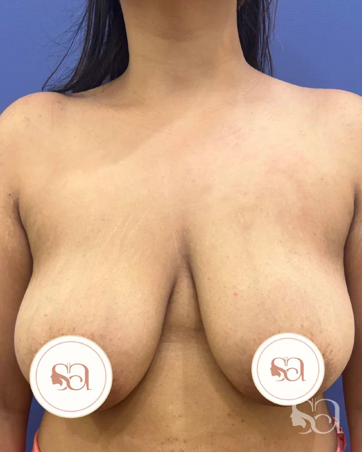 Before
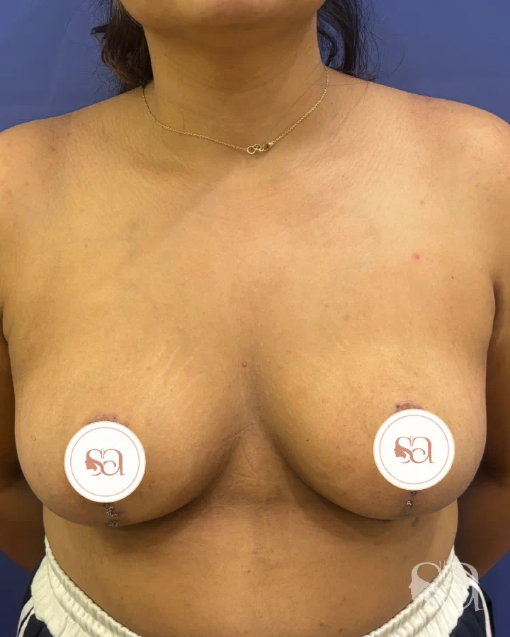 After
AfterThis result shows an unclothed body after surgery.
Breast Reduction, case 1
- 8 weeks after surgery
- Results vary between individuals
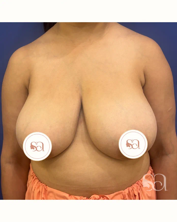 Before
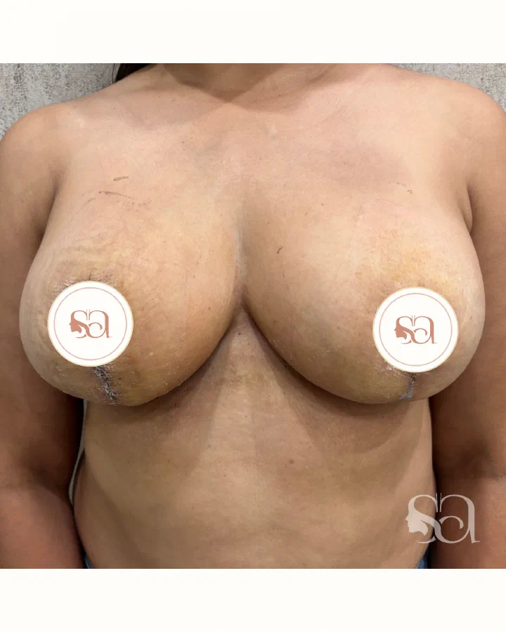 After
AfterThis result shows an unclothed body after surgery.
Breast Reduction, case 2
- 3 weeks after surgery
- Results vary between individuals
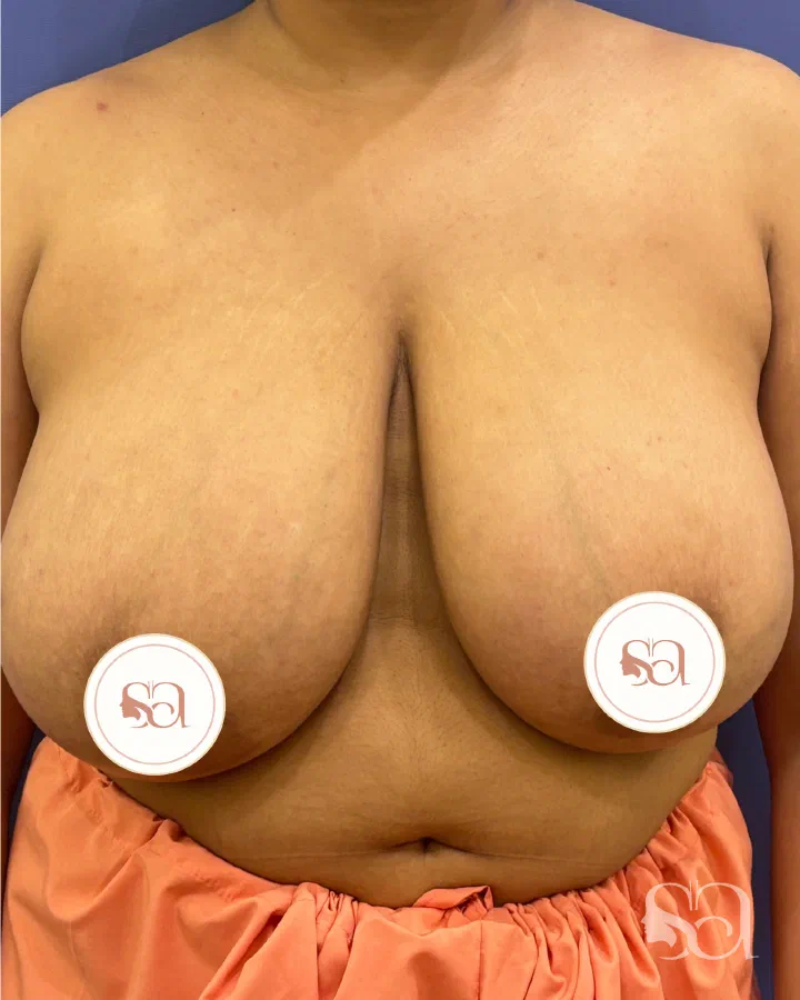 Before
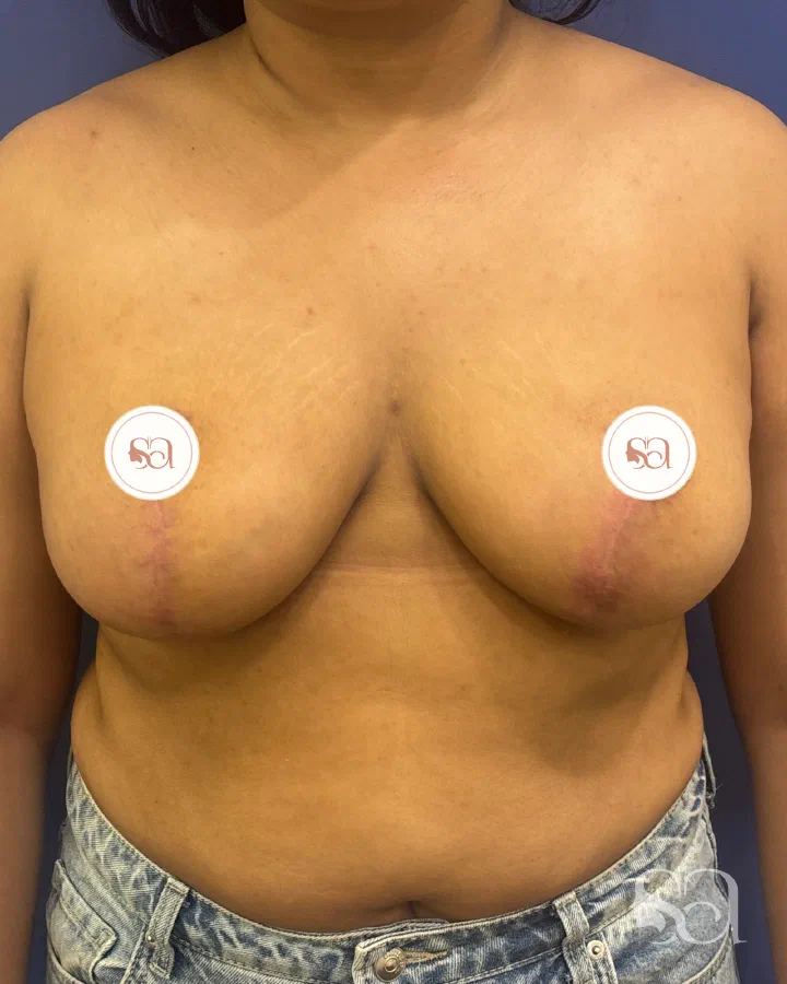 After
AfterThis result shows an unclothed body after surgery.
Breast Reduction, case 3
- 3 months after surgery
- Results vary between individuals
Photographs of the clinic's own patients, shown with their written consent, with faces blurred or covered as far as the result allows. They explain what an operation can change; they are not a promise of your result.
Which reduction technique fits breast size, droop, and tissue type
Reduction mammoplasty is not one fixed operation. The technique is selected by reduction volume, ptosis, breast base width, skin quality, areola size, tissue density, scar tolerance, nipple-sensation concerns, and future breastfeeding priorities.
Two separate decisions are made, and they are not alternatives to each other. The first is the pedicle - the bridge of tissue that keeps the nipple-areola complex alive while the surrounding gland, fat, and skin are removed. The second is the skin pattern - where the scars sit. Almost every excisional reduction needs one of each, and the combination most commonly performed is an inferior pedicle with an anchor-pattern skin excision, so a patient is rarely choosing between those two.
The pedicle options:
Inferior pedicle keeps the nipple-areola complex attached to a lower bridge of breast tissue while tissue is removed around it. It is reliable for moderate to large reductions, heavier breasts, and cases where preserving nipple blood supply is the main safety priority.
Superomedial or superior pedicle keeps the nipple attached to tissue from the inner or upper breast. It can give good upper-pole shape in moderate reductions where the nipple does not need an extreme lift.
When transposition on any pedicle would be too long to be safe - usually in very large reductions - a free-nipple graft is discussed instead, with the trade-offs set out in the size question further down this page.
The skin-pattern options:
Vertical or lollipop pattern places a scar around the areola and down to the crease, usually without the horizontal fold scar. It suits moderate reductions with less lower-pole skin excess and better skin recoil.
Wise-pattern or anchor adds a horizontal scar hidden in the breast fold to the areolar and vertical scars. It is chosen when the breast is heavy, the lower pole is stretched, the nipple sits far below the crease, or the skin envelope needs powerful tightening. Because this is the pattern most often paired with an inferior pedicle, a patient planned for an inferior-pedicle reduction should expect the in-fold scar unless told otherwise at marking.
Separate from both of those axes, liposuction-only reduction removes fat through small cannula entries but does not lift the nipple or remove loose skin, and because the tissue is aspirated rather than cut out, no specimen is available for pathology the way it is after an excisional reduction. It is not an excisional reduction and is reserved for fatty breasts with good skin elasticity, minimal ptosis, and no need for areola repositioning.
Breast reduction almost always contains a lift component because the nipple is repositioned and the breast mound is reshaped. The overlap with breast lift surgery in Gurgaon is discussed openly: lift controls position and skin, while reduction adds meaningful tissue and weight removal.
Areola size, nipple position, and how much of that a reduction changes
A large areola is normal anatomical variation, not a disease. Diameter is largely genetic to begin with, and it is then stretched by breast growth in puberty, pregnancy, breastfeeding, and weight change. Once that skin has stretched, no cream, oil, massage, ice pack, or exercise shrinks the areola border. Areola diameter is changed surgically or it is not changed at all.
Because every reduction pattern cuts around the areola border, areola reduction is built into the operation rather than added to it. The new diameter is marked with a circular template while the patient is standing, chosen for the patient’s breast base width and frame, and the surrounding skin is then gathered in to that smaller circle. Patients who came in about breast weight often notice this change most in the first month, before swelling settles and the lower pole finds its shape.
A standalone periareolar areola reduction - a ring of skin only, with no tissue removal and no lift - is possible when volume and nipple height are already acceptable, and it can be done under local anaesthesia or sedation. The trade-off is stated before it is offered: the scar sits on the areola border under tension, so it can widen, fade unevenly, or flatten areola projection. When the nipple also sits low, a breast lift that includes areola reduction usually holds shape better than periareolar excision alone.
Asymmetric areolae are marked per side. Nipple height, areola diameter, and skin removal are set independently on the left and the right. Visible imbalance is usually improved, but exact symmetry is not promised, because the rib cage and the breast bases underneath differ from one side to the other.
Talk it through with Dr. Shikha
Book a consultation at Dr. Shikha Aesthetics in Sushant Lok 1. Bring your questions about breast reduction, and any previous reports.
How the plan is matched to symptoms, cup goals, and breastfeeding priorities
The first planning question is what problem the patient needs solved. A patient with shoulder grooves, chronic rashes, and a very heavy lower pole needs a different operation from a patient who wants a one-cup reduction for clothing fit. The amount removed is estimated from breast volume, cup-size goal, body frame, and symptom burden; the final size is planned by proportion and safe tissue handling, not by promising an exact bra cup.
Ptosis grade is measured standing. If the nipple sits near the breast fold and the skin is firm, a smaller vertical pattern may be enough. If the nipple sits well below the fold, points downward, or the lower breast hangs heavily against the chest wall, an anchor reduction is more likely. If one breast is larger or lower, each side may need a different amount removed and a different skin pattern to improve symmetry.
Tissue type changes the plan. Dense glandular breasts usually need direct removal, while fatty breasts with good recoil may accept liposuction as an adjunct for side fullness. Thin, stretch-marked skin is treated cautiously because it can stretch again.
Future breastfeeding is discussed before the scar pattern is chosen. Modern pedicle techniques aim to preserve blood supply, nerves, and some duct connections to the nipple-areola complex, but breastfeeding cannot be guaranteed after any reduction. Risk rises when very large volumes are removed, the nipple moves a long distance, or a free-nipple graft is considered.
For patients trying to understand lactation after previous or planned breast surgery, the clinic links the surgical discussion to the companion guide on breastfeeding after breast surgery in India rather than duplicating that full topic on this procedure page.
Dr. Shikha also references the candidate-selection video on this page because symptom triggers matter: the examination answers whether breast weight justifies scars, anaesthesia, and recovery.
What happens from consultation to same-day discharge
The first consultation usually takes 30 to 45 minutes. The history covers pain pattern, shoulder grooves, rashes, exercise restriction, previous pregnancies, breastfeeding history, future pregnancy plans, weight stability, medications, nicotine use, diabetes, family history of breast cancer, and previous breast imaging or surgery. The examination documents breast size, asymmetry, nipple height, areola diameter, skin quality, side fullness, fold position, and lift requirement.
Standard photographs and standing measurements are taken for planning. Mammography is obtained for patients aged 40 and above and for patients with a family history of breast cancer. Ultrasound or further imaging is added at any age when there is a lump, nipple discharge, or a previous breast concern. Routine blood tests, ECG, and anaesthesia fitness are completed before the date is confirmed.
The operation is performed under general anaesthesia in an accredited day-care operating setup. Markings are made standing before anaesthesia. A typical reduction takes three to four hours; very large reductions, major asymmetry, or combined procedures take longer. Excess tissue is removed, the breast is reshaped internally, the nipple-areola complex is moved on its pedicle, and the skin is closed in layers.
Drains are not automatic but may be used when tissue removal is large. A support bra is applied before discharge. Most primary reductions are discharged the same day or after overnight observation depending on operative duration, drain use, comfort, and anaesthesia recovery.
Risks and complications to weigh before deciding
Reduction mammoplasty is a well-studied operation with high satisfaction, but it is real surgery on a tissue flap that carries its own blood supply, and the complication profile is put in front of the patient before consent rather than after.
Wound healing and the inverted-T junction. Delayed healing and wound separation are the commonest problems after a reduction, and the usual site is the inverted-T junction where the vertical and horizontal scars meet in an anchor pattern - the point under the most tension with the least blood supply. Most settle with dressings and time; a minority need a small secondary closure or a scar revision once healed.
Bleeding and haematoma. Bleeding into the breast in the first 24 to 48 hours can collect as a haematoma, and this is the commonest complication serious enough to need a return to theatre for drainage. Sudden one-sided swelling, tightening, or disproportionate pain is reviewed urgently rather than watched.
Infection and seroma. Cellulitis or a wound infection is treated with antibiotics; a fluid collection under the flap sometimes needs aspiration. Drains are used at the surgeon’s discretion after a large resection, and trial evidence has not shown that they reduce seroma or haematoma.
Fat necrosis. A firm, tender lump can form where a patch of fat loses its blood supply. It usually softens over months and is managed conservatively, but it can leave a palpable area, produce a small discharge, or appear on later imaging and need to be distinguished from an unrelated breast lump.
Nipple-areola perfusion. Because the nipple-areola complex survives on its pedicle, partial loss of areola skin - and, rarely, loss of the whole complex - is possible. Congestion or colour change in the first hours is treated as an emergency rather than a wait-and-see finding. This is also the reason a free-nipple graft is discussed instead when the requested reduction is extreme or the nipple has to travel a very long distance.
Venous thromboembolism. A general anaesthetic followed by immobility carries a small risk of deep vein thrombosis and pulmonary embolism. That risk rises with longer theatre time, higher BMI, and combining the reduction with abdominal or body contouring in one sitting. Early walking, calf exercises, hydration, and compression are used routinely, and any calf pain, one-sided leg swelling, breathlessness, or chest pain after surgery is treated as urgent.
What raises these risks. Nicotine in any form, a BMI of 30 or above, diabetes, previous chest-wall radiation, very large resection volumes, and long nipple transposition all push wound-healing and blood-supply risk upward - the same modifiers listed later for nipple sensation apply to healing and perfusion first. This is why nicotine cessation for at least four weeks either side of surgery is treated as non-negotiable rather than as advice.
None of this argues against a reduction when breast weight is genuinely causing symptoms. It is the reason the operation is planned around measurements, tissue handling, and anaesthetic fitness rather than a target cup size, and each of these points is worked through in person before consent.
What recovery looks like after breast reduction
Breast reduction recovery is summarized here because the detailed diary belongs in the companion guide on week-by-week breast reduction recovery.
Week 0 to 1: swelling, tightness, bruising, and moderate soreness are expected. The support bra stays on day and night. Arm movement is gentle, and lifting children, pushing heavy doors, overhead reaching, stomach sleeping, driving, and household work are avoided. The first review is commonly around day 5 to 7 for dressing check and drain removal if drains were used.
Week 2: many patients return to desk work when pain medicine is no longer sedating and arm movement is comfortable. The breasts may look high, swollen, or boxy at this stage; this is normal early settling, not the final shape.
Weeks 3 to 4: walking and light daily tasks feel easier. Minor pulling, side tightness, reduced nipple sensation, or small numb patches are common while nerves recover. Scar care may begin once wounds are fully closed.
Weeks 5 to 6: light cardio is often reintroduced after review. Running, upper-body weights, swimming, yoga inversions, and chest-loading exercise wait until the wounds are mature enough and surgeon clearance is given.
Month 3 onward: swelling has reduced enough for a meaningful shape review. Scars keep maturing for 12 to 18 months, and lower-pole settling continues gradually across the first year.
Why results change with weight, pregnancy, and time
Breast reduction gives a long-lasting reduction because removed tissue does not grow back in the same way. It does not freeze the breast against ageing, gravity, hormones, pregnancy, breastfeeding, or major weight change. Stable weight is one of the strongest predictors of durability.
Pregnancy after reduction can stretch the skin envelope, enlarge the breast during pregnancy, reduce volume after weaning, and change nipple position. It does not make pregnancy unsafe, but it can shorten result durability.
Nipple sensation may decrease, increase, or feel patchy after surgery. Many changes improve over months, but permanent sensation change is possible, especially after larger reductions, long nipple movement, previous surgery, diabetes, nicotine exposure, or free-nipple graft planning.
Scars are permanent. A vertical reduction leaves a scar around the areola and down to the breast crease; an anchor reduction adds a scar hidden in the fold. Scars are typically pink and firm for three to six months, then soften and fade over 12 to 18 months.
Revision is considered when a scar widens, a dog-ear remains, asymmetry persists after swelling settles, the areola stretches, or major weight or pregnancy change creates new looseness. Small scar revisions are usually assessed after scars mature, not in the first few weeks.
Breast reduction in Gurgaon and Delhi NCR - what patients should plan around
The clinic sees breast reduction patients from Gurgaon, Delhi, Noida, Faridabad, Ghaziabad, and other parts of Delhi NCR. Common consultation stories include back and shoulder discomfort, humid-season rashes, difficulty finding supportive bras, gym avoidance, desk-job posture strain, and the wish to look proportionate without becoming too small.
Consultations are appointment-led because planning requires measurements, standing assessment, and a direct discussion about scars, cup-size expectations, nipple position, breastfeeding, and recovery logistics. Out-of-NCR patients should usually stay 7 to 10 days for the first dressing review and wound check.
Desk-based patients often arrange 10 to 14 days away from office pressure, while field work, childcare lifting, healthcare work, or travel-heavy jobs need longer support.
Breast reduction can also be part of a wider post-pregnancy plan when abdominal laxity, diastasis, or stubborn fat areas are part of the same consultation. Combined surgery is planned around BMI, anaesthesia duration, haemoglobin, diabetes control, childcare support, and recovery capacity - those inputs matter because a single longer anaesthetic and a larger immobile recovery raise venous thromboembolism (deep vein thrombosis and pulmonary embolism) and wound-healing risk compared with staging the two operations. The broader safety logic is covered on the mommy makeover surgery in Gurgaon page.