
Rhinoplasty reshapes the bone, cartilage and, where needed, soft tissue of the nose to refine the dorsum, tip, alar base, or breathing airway - through an open or closed approach, with or without a septoplasty component for combined functional repair. At Dr. Shikha Bansal’s clinic in Gurgaon the technique is matched to the individual nose at consultation rather than chosen by default: features common in Indian noses (thicker tip skin, softer lower lateral cartilages and a broader alar base, though anatomy varies widely) change the planning compared with the Caucasian template that most global content is written around.
Key points
- Open, closed, septorhinoplasty, and revision approaches matched to the specific nose
- Planning for features common in Indian noses (thicker skin, softer tip cartilage, broader alar base), where present
- Structural grafting (septal, conchal, or rib cartilage) used when projection or definition needs support
- Ultrasonic (piezo) instrumentation when the planning warrants it
- Same-day discharge for most patients, with structured Day 0 to Month 18 recovery counselling that includes the long Indian-tissue settling tail
- Liquid rhinoplasty not offered - patients who specifically want fillers are referred to a qualified dermatologist or aesthetic physician
What rhinoplasty can address, and what it cannot
Rhinoplasty - surgical reshaping of the nose - is the right call when a specific anatomical feature is genuinely unbalanced for the rest of the face, when a functional breathing problem coexists with a cosmetic concern, or when an earlier rhinoplasty needs corrective work. It is a poor call when the patient is searching for a face the nose cannot give them, when the dominant concern is actually skin or pigment rather than shape, or when realistic expectations have not settled.
The well-defined problems rhinoplasty addresses are a dorsal hump, a wide or bulbous tip, a drooping or under-projected tip, a wide alar base, a deviated nose or nasal bone, a deviated septum that blocks airflow, post-traumatic deformity, and outcomes from a previous nasal surgery that did not settle as planned. Each of these has a different surgical plan; lumping them together as “a nose job” loses the precision that makes a good result possible.
Rhinoplasty does not change skin texture, treat acne or pore size on the nose, lighten pigmentation, or alter facial expression beyond what the nose itself contributes. It also does not narrow a thick-skinned nose to a thin-skinned look - the soft tissue envelope is the patient’s own skin and it determines how much definition can show through, no matter what the underlying cartilage is reshaped to. This is an especially important conversation in Indian rhinoplasty because tip definition lives behind the skin, and patients who arrive with an image of a thin-skinned celebrity nose need to know what the skin envelope will and will not allow.
Surgical rhinoplasty is also not the same as filler-based “liquid rhinoplasty”. The clinic does not offer liquid rhinoplasty. The honest position on fillers in the nose is covered separately on the liquid rhinoplasty guide: a small set of features can be camouflaged temporarily with hyaluronic-acid filler placed by a trained dermatologist or aesthetic physician, but the nasal vascular anatomy is unforgiving and filler complications in the nose are among the most serious in cosmetic medicine. Patients who want a durable structural change are assessed for surgery at consultation; patients who specifically want fillers are sent to a qualified dermatologist or aesthetic physician rather than treated at this clinic.
Rhinoplasty techniques - open, closed, septorhinoplasty, and ethnic considerations
There is no single best rhinoplasty technique. The choice runs across several decisions, each with a clean clinical rationale rather than a one-size answer. The vocabulary patients arrive with - open versus closed, primary versus revision, cosmetic versus functional - maps onto an internal decision set that the surgeon walks through at consultation.
Open rhinoplasty uses a small bridging incision across the columella (the strip of skin between the nostrils) connecting to incisions inside the rim of each nostril. Once the skin envelope is lifted, the underlying cartilage and bone are visible to both eyes simultaneously and instrumented under direct vision. The trade-off is a small external scar at the columella that fades to a fine line and a slightly longer settling timeline at the tip. Open is the default at the clinic for complex tip work, secondary or revision cases, septorhinoplasty with structural grafting, ethnic noses requiring tip refinement on a thicker skin envelope, and any case where intra-operative judgment depends on seeing the architecture directly. The columellar scar is one of the most common patient anxieties at consultation and in most patients is hard to see by six months, though in darker skin it can take longer to fade.
Closed (endonasal) rhinoplasty keeps every incision inside the nostrils. There is no external scar unless an alar base reduction is added, which leaves a small scar in the nostril crease. The surgeon works through a smaller working space with reduced direct vision. It is the right choice for selected dorsal-only work - straightforward hump reduction in a patient with sound tip support and a strong septum - and for very limited tip-only refinements. It is generally less suited to substantial tip restructuring, revision rhinoplasty, or ethnic noses where grafts are commonly needed.
Septorhinoplasty combines septoplasty (straightening the internal nasal septum to restore the breathing airway) with rhinoplasty (changing the external shape). It is the right plan when a patient has a deviated septum and is unhappy with how the nose looks - both problems are addressed in a single anaesthetic. The functional and cosmetic components are planned and discussed separately. The detailed pathway is covered on the septorhinoplasty guide.
Cartilage grafting is a routine part of rhinoplasty rather than an exotic add-on. Septal cartilage is the first source - harvested from the same septum during septoplasty. Ear (conchal) cartilage is a second source when more material is needed. Rib cartilage is used mainly for revision rhinoplasty, severe traumatic deformity, substantial structural rebuild, or significant dorsal augmentation in a first operation, where it provides ample, strong graft material. Rib cartilage harvest adds a chest scar and donor-site discomfort, and carries a small risk of graft warping and, rarely, pneumothorax; these are discussed at consultation. Indian noses needing tip projection or dorsal augmentation often need grafting on the primary; this is planned at consultation rather than discovered intra-operatively.
Preservation rhinoplasty is a revived approach that preserves the existing dorsal aesthetic line by lowering the entire dorsum from below rather than rasping or removing the hump from above. In carefully selected patients it can give a smooth dorsal line; residual or recurrent hump is a recognised limitation, and long-term evidence is still maturing. It is not a replacement for traditional structural rhinoplasty in every case - patient anatomy and the specific problem decide.
Ultrasonic (piezo) instrumentation uses a piezoelectric handpiece to sculpt the bony pyramid with millimetric control with less trauma to the surrounding soft tissue. Studies suggest it can reduce early swelling and bruising compared with osteotomes (small surgical chisels) in selected cases. The clinic uses piezo instrumentation when the planning warrants it; not every nose needs it. A detailed view of where modern instrumentation actually changes outcomes is covered on the ultrasonic rhinoplasty guide.
Ethnic and Indian-anatomy considerations sit through every one of the decisions above. Many Indian noses, though anatomy varies widely across regions, have a thicker tip skin envelope, softer lower lateral cartilages that need structural support, a broader alar base and, in some patients, a lower dorsum than the Caucasian template that most surgical training literature was written for. The implications are practical: more grafting on the primary, alar base reduction more frequently considered, longer tip settling because thick skin remodels slowly, and a more conservative dorsal reduction so the nose does not end up scooped in a way that does not suit Indian facial proportions. The Indian nose anatomy and ethnic rhinoplasty planning guide walks through this in detail.
How the technique is matched to the patient
At the consultation the nose is examined in six standard views - frontal, three-quarter left and right, profile left and right, and basal - under standardised lighting. The skin envelope is pinched at the supratip to estimate thickness; the lower lateral cartilages are palpated for strength and width; the septum is examined with a nasal speculum for deviation, perforation, or prior surgical alteration. The patient’s history is reviewed for nasal trauma, prior nasal surgery, allergic rhinitis, smoking, bleeding disorders, and any medications that thin the blood. A 3D imaging session may be added for selected patients, with the explicit caveat that the simulated image is a planning aid and not a guarantee - the actual healing of the skin envelope on the new framework is what produces the final result.
Dorsal hump dominant - the surgical plan is dorsal reduction (rasp, osteotome, or piezo for the bony portion; sharp resection or component reduction for the cartilage portion) with paired lateral osteotomies to narrow the residual bony base. Open or closed approach depends on whether tip work is added. Preservation rhinoplasty is considered when the dorsal aesthetic line is good and only height needs to come down.
Bulbous or under-projected tip - tip refinement through cartilage suturing techniques (dome suturing, transdomal sutures, interdomal sutures) supported by structural grafts (columellar strut, septal extension graft, or shield graft) to project and define. Indian noses with thick skin and weak lower lateral cartilages often need a structural graft to hold the new shape against the recoil of the skin envelope.
Drooping tip - a septal extension graft or columellar strut to support the tip and rotate it upward, with tip suturing to refine. Patients whose tip drops on smiling may have an overactive depressor septi nasi muscle, which can be released in the same operation.
Short or over-rotated nose - a septal extension graft to lengthen and derotate the tip downward.
Deviated nose with breathing obstruction - septorhinoplasty with septal straightening, spreader grafts to open the internal nasal valve, paired osteotomies to realign the external pyramid, and tip work as needed. Functional and cosmetic are planned together to avoid two operations.
Wide alar base - alar base reduction (Weir excision or sill excision) brings the nostril width in. The decision to add alar base reduction is made cautiously because the Weir scar sits in the natural alar crease (a sill excision scar sits at the nostril sill) and an over-aggressive reduction produces a pinched, unnatural look. In Indian patients with wide bases the planning is conservative and staged: small reduction first, more later only if needed.
Revision after a previous rhinoplasty - the plan is different from primary work because tissue planes are altered, cartilage support may be depleted, and the skin envelope’s healing behaviour has changed. The open approach is the default, rib cartilage may be needed for structural rebuild, and the case is planned with a clear understanding that the result of a revision is constrained by what the previous surgery left to work with. The detailed pathway is covered on the revision rhinoplasty guide.
Post-traumatic deformity - staged or single-stage repair depending on how long ago the trauma occurred, whether the septum is involved, and whether breathing has been compromised. A nose that was broken and reset incompletely a few years ago has different planning needs from a fresh fracture.
Patient priorities and what the nose cannot give - patients whose primary motivation is a celebrity-image match are walked through the limits of their own skin envelope and facial proportion at consultation. A nose can be improved within the patient’s own architecture; it cannot be replaced with someone else’s. This conversation is held up-front because expectation alignment is one of the single largest predictors of patient satisfaction in rhinoplasty literature.
Talk it through with Dr. Shikha
Book a consultation at Dr. Shikha Aesthetics in Sushant Lok 1. Bring your questions about rhinoplasty, and any previous reports.
The procedure, from consultation to splint removal
The first consultation runs 45 to 60 minutes. The nose is examined in the six standard views, the septum is checked internally, photographs are taken in standardised lighting, and the patient is asked to bring or describe the look they are working toward. Imaging - clinical photographs at minimum, 3D simulation in selected cases - supports the conversation about achievable change. A pre-operative ENT review is added when the patient reports breathing obstruction, recurrent sinusitis, or known septal deviation.
Pre-operative work-up includes a baseline blood profile, ECG and physician fitness clearance, and instructions to pause blood thinners only on the advice of the doctor who prescribed them, to stop aspirin, anti-inflammatory painkillers and supplements such as fish oil, vitamin E and ginkgo as instructed, to pause oestrogen-based medications where the physician advises, and to stop smoking for at least four weeks before and four weeks after surgery. Smoking significantly raises the risk of skin-edge breakdown and poor cartilage healing in rhinoplasty, so stopping it is non-negotiable - the soft tissue at the columellar incision is particularly vulnerable, and a smoker is at meaningfully higher risk of a poor scar or skin-edge complication.
The operation is performed under general anaesthesia in a day-care operating theatre, with a qualified anaesthesiologist managing the airway. Surgical time is typically 90 to 180 minutes for a primary cosmetic rhinoplasty, longer for septorhinoplasty with structural grafting and longer again for revision work. The chosen incisions are made (a columellar incision plus incisions inside the nostril rims for open; endonasal only for closed), the skin envelope is lifted in the correct plane, the bony and cartilage framework is reshaped, septal cartilage is harvested if grafting is planned, osteotomies are performed when needed, the grafts are placed and sutured into the planned positions, and the skin envelope is redraped. The incisions are closed with fine absorbable sutures and one or two non-absorbable sutures at the columella that are removed at the first follow-up. An external nasal splint is applied - moulded to the new dorsum - and internal silicone splints are placed inside each nostril if the septum was operated on. A small mustache dressing under the nose catches the small amount of drainage that is normal in the first 24 hours.
Most patients are discharged the same day, three to five hours after the surgery ends, once they are alert, can drink fluids, and have walked to the bathroom independently. Overnight observation is offered when the patient lives more than 90 minutes from the clinic, when the surgery was extended, or when a comorbidity warrants it. A family member drives the patient home with the head propped up.
Recovery after rhinoplasty, week by week
Rhinoplasty recovery follows a long but predictable arc. The visible part - bruising, splint removal, the immediate post-splint nose - runs over the first three weeks. The invisible part - internal tip and supratip swelling resolving as the skin envelope settles onto the new framework - runs over twelve to eighteen months in thicker-skinned noses, which are common in Indian patients, longer than the often-quoted twelve months that comes from Caucasian-tissue literature. Patients are told this at consultation rather than discovered at month six.
Day 0 to Day 3 - bruising around the eyes is common and peaks at day two to three before fading. Mild oozing from the nostrils on to the mustache dressing is normal and is changed as needed. The internal silicone splints (if placed for septoplasty) make the nose feel blocked, like a head cold, so most patients breathe mainly through the mouth until they are removed. Sleeping propped up at 30 to 45 degrees helps reduce swelling. Cold compresses are used around the eyes (never directly on the nose) in 15-minute intervals for the first 48 hours. Pain is usually mild to moderate and is managed with oral painkillers, and many patients find the blocked-nose feeling more bothersome than the pain; opioids are rarely required. If rib cartilage is harvested, the chest donor site is usually more uncomfortable than the nose for the first one to two weeks.
Day 4 to Day 7 - bruising starts to fade through the typical yellow-green resolution sequence. The internal silicone splints are removed at day five to seven at the first follow-up; mouth-breathing eases dramatically once they are out. The external splint stays on. Patients with a sedentary job typically return to work between day seven and day ten, ideally after the external splint comes off and surface bruising has faded enough for makeup coverage. Driving resumes once the patient is off any sedating painkillers, and not within 48 hours of the anaesthetic.
Week 2 - the external splint is removed at the day seven to ten visit. The nose appearing immediately after splint removal is not the final result - it is swollen, the bridge looks higher than it will end at, and the tip looks fuller. Patients who are not warned about this find the splint-off visit emotionally difficult; patients who are warned recognise it as the expected stage. Light walking and gentle activity are fine; nothing that risks a knock to the nose, no contact sports, no children-on-shoulders moments. Glasses cannot rest on the nasal bridge for six weeks - taped to the forehead or contact lenses are the work-arounds.
Weeks 3 to 6 - surface bruising fully resolved. The bony pyramid has begun to consolidate but is not yet fully stable; nasal trauma in this window can displace the bones. Light cardio resumes from week three; weights, core and other strenuous gym work from around week six, or as the surgeon advises; swimming with goggles pressing on the bridge and any movement that risks a direct hit to the nose are held back for at least six weeks, and contact sports where a blow to the nose is likely for around three months, or as the surgeon advises.
Month 3 - the obvious swelling has resolved enough that the nose looks largely like the planned result in clothing, photographs from a distance, and most social settings. Up close, the tip is still firmer than it will become, and small contour irregularities at the supratip may still be present. The bony pyramid is stable. Many patients find the nose looks close to the planned result in everyday settings at this stage.
Months 6 to 12 - fine-tuning of swelling continues, particularly at the tip and supratip. Patients with thicker skin (Indian, Middle Eastern, North African anatomical patterns) take longer at this stage than patients with thin skin. Tip definition continues to emerge through this window.
Month 12 to Month 18 - final settling in patients with thicker skin. The full result is judged at twelve to eighteen months, not at three months and not at six. Patients considering whether a small refinement might be warranted are advised to wait until the full settling has occurred before a touch-up is discussed; the nose at six months will not look the same as the nose at fifteen months, and an early revision decision is often premature because the nose is still changing.
A more granular week-by-week recovery view - splint changes, social return timing, makeup application, and bridge-of-glasses workarounds - is covered on the rhinoplasty recovery week by week guide.
Complications, revision, and long-term results
Rhinoplasty is one of the most technically demanding operations in plastic surgery because the result lives in millimetric changes to a structure that is visible from every angle and used every day for breathing. Most rhinoplasties heal uneventfully; the realistic conversation at consultation covers the small set of complications that are worth knowing about and how the clinic minimises and manages them.
Bruising and prolonged swelling are common early and resolve over the first three weeks at the surface and over twelve to eighteen months internally. Persistent tip swelling beyond what the patient expects is a common source of post-operative dissatisfaction in Indian rhinoplasty and is more often a settling-timeline issue than a true complication. Patient education at consultation about the long settling tail is more useful than any post-operative intervention.
Bleeding in the first 24 to 48 hours is the most common acute complication. Small amounts of oozing on the mustache dressing are expected; brisk bleeding that soaks through repeatedly is not, and is the trigger for a call to the clinic. Bleeding risk is reduced by managing blood thinners before surgery on the prescribing doctor’s advice and controlling blood pressure intra-operatively. When the septum has been operated on, internal silicone splints support it and reduce the risk of septal haematoma and adhesions.
Infection is uncommon in rhinoplasty because the nose has an excellent blood supply. Routine peri-operative antibiotics are used. A persistent unilateral discharge or worsening pain beyond the first week warrants review.
Other risks include septal haematoma or perforation, numbness of the tip and upper lip (usually settling over months, occasionally persistent), a change in sense of smell (usually temporary), nasal adhesions, rarely toxic shock syndrome from internal splints or a saddle-nose collapse after septal work, and the general risks of anaesthesia. Increasing nasal blockage with pain after septal work, or fresh bleeding in the first two weeks, needs same-day review. Revision rates for primary rhinoplasty are commonly quoted at about 5-15% in published series.
Asymmetry and contour irregularities are the small-deviation outcomes that drive most revision conversations. Causes include uneven healing of the bony pyramid after osteotomies, supratip fullness from incomplete cartilage reduction, tip asymmetry from uneven graft positioning, and small dorsal irregularities that show through thin skin. Most are minor; some can be addressed with closed touch-ups at twelve to eighteen months once full settling has occurred.
Breathing changes can run either direction. Septorhinoplasty usually improves airflow when a deviated septum is the main cause of obstruction, though turbinate swelling or allergic rhinitis can still cause some blockage afterwards; cosmetic rhinoplasty done without attention to the internal nasal valve can narrow it and produce a sense of obstruction that was not present before. The clinic’s planning aims to protect the internal nasal valve (spreader grafts are placed when narrowing risk is anticipated) to reduce the risk that reshaping narrows the airway.
Skin envelope complications - the columellar incision and the soft tissue overlying the new framework are vulnerable to vascular compromise in smokers and in patients with very tight skin redraping after large reductions. Smoking cessation for the recommended window is the single largest controllable factor.
Long-term changes to settling - the nose continues to remodel for years. Cartilage memory, scar contracture, and skin envelope behaviour all contribute. A nose that looks excellent at year one may look slightly different at year five; in most patients the changes are subtle and within the planning envelope. In some patients a small revision becomes worth considering after full settling.
Revision rhinoplasty is a separate operation from the primary, planned on its own. The right time to consider revision is after the nose has fully settled - twelve to eighteen months for thicker skin, longer in some patients - and after a clear conversation about what the revision can and cannot change given the tissue available to work with. The pathway is covered on the revision rhinoplasty guide. The honest separation of what is normal healing from what is a true complication is the focus of the rhinoplasty side effects and complications guide.
Rhinoplasty in Gurgaon and Delhi NCR - what to expect
The clinic sees rhinoplasty patients from across Delhi NCR - Gurgaon, Delhi, Noida, Faridabad, Ghaziabad - and selected patients from outside the region. Most patients come for one or two consultations before surgery; rhinoplasty is one of the procedures least suited to a same-day consultation-and-decision visit, and patients are encouraged to live with their plan for a few weeks before committing.
The clinic is in Sushant Lok 1, Gurgaon. Out-of-station patients are advised to plan a stay of approximately ten days post-operatively before flying home - the splint comes off at day seven to ten and travel is more comfortable with surface bruising mostly faded. Flying is usually comfortable after the splint-removal visit; patients are advised on hydration, moving during long flights, and nasal care for the journey. The practical reason for the stay is the splint-removal visit and the early follow-up window.
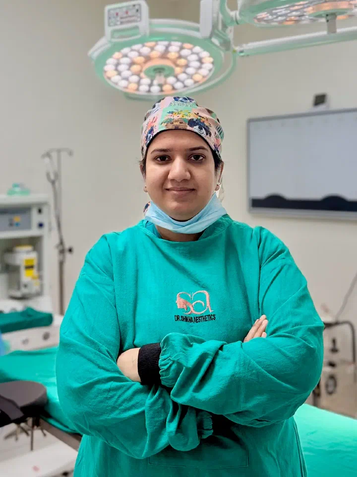
Consultations are by appointment. Photographs taken at consultation are stored securely and used only for clinical planning unless the patient explicitly consents in writing to before-and-after use for educational purposes. The clinic’s positioning on rhinoplasty is surgical - open and closed, primary and revision, cosmetic and septorhinoplasty - and non-surgical “liquid rhinoplasty” with filler is not offered. Patients who specifically want a temporary filler-based change are referred to a qualified dermatologist or aesthetic physician for that conversation rather than treated at this clinic, and that referral is offered honestly rather than as a disparaging comment about the underlying treatment. Dr. Shikha Bansal’s credentials - MCh Plastic and Reconstructive Surgery, Haryana Medical Council registration 24859, Ex-Fellow at Artemis Hospital’s Cosmetology and Plastic Surgery Centre, and active membership of IAAPS and APSI - are the same on this page as on the clinic’s about page so a patient can verify them independently before booking.